
Most people injured in a collision do not think about testing for days. They go home, wait to feel better, and start looking for answers only when the fog does not lift.
By then some of the best evidence has already gone. Not because anyone disbelieved them — because certain markers physically clear from the blood on a schedule, and that schedule does not wait for anyone to decide the symptoms are serious enough.
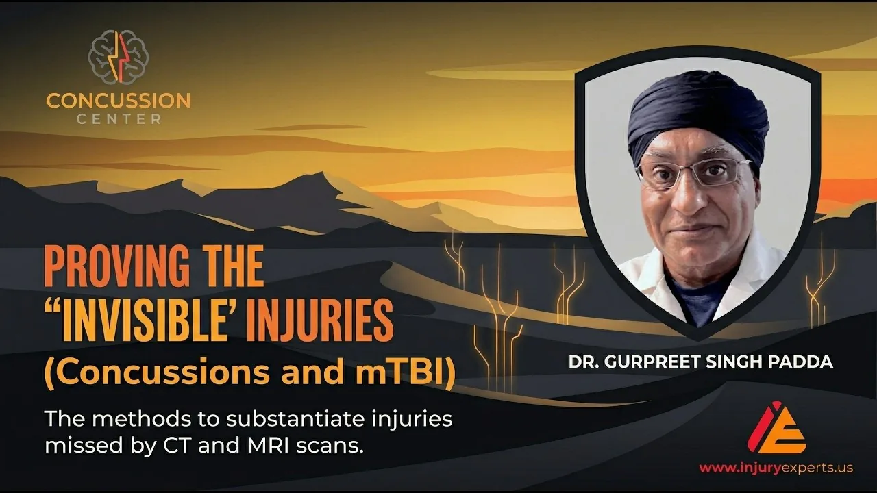
Watch on YouTube: Proving the Invisible & Architecting Regeneration
Structural damage versus functional impairment
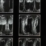
CT and MRI answer one question well: is there a fracture or a bleed? Mild traumatic brain injury is primarily a microscopic event — axonal shearing and a metabolic mismatch in which injured tissue cannot get the energy it needs to repair. Standard neuroimaging is not built to detect that.
This gap is where claims are denied: if it is not on the scan, it is not real. Two categories of testing close the gap.
The blood markers, and their windows
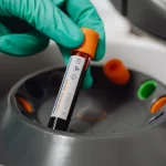
Since 2018, blood biomarkers have made cellular distress measurable. Two matter most acutely, and their timing differs in a way that changes what you should do:
| Marker | Source | Rises | Peak | Useful window |
|---|---|---|---|---|
| UCH-L1 | neuronal cell bodies | rapidly | ~8 hours | declines rapidly over ~48 hours |
| GFAP | astrocytes (glial) | rapidly | ~20 hours | declines slowly over ~72 hours; performs across 7 days |
The practical consequence is stark. UCH-L1 is a hyperacute signal — wait two days and the window has closed. GFAP is the more forgiving marker and remains useful for people who seek care later in the week.
The combined GFAP/UCH-L1 panel is used chiefly as a rule-out, and here the evidence is strong. The ALERT-TBI study (Bazarian et al., The Lancet Neurology, 2018) reported sensitivity of 0.976 and a negative predictive value of 0.996 for traumatic intracranial abnormalities on CT. The authors estimated that routine use could reduce CT scans by roughly a third.
Read that correctly, because it is often misread. A very high negative predictive value means a negative result is strong evidence against the injury being screened for. It does not mean a positive result proves a specific diagnosis or predicts your recovery.
Point-of-care platforms can return these results within roughly fifteen minutes, which is what makes them useful for an immediate decision about whether a radiation-heavy CT is warranted at all.
Verified against primary sources. The timing figures come from Papa et al., JAMA Neurology (2016); the 0.996 negative predictive value from Bazarian et al., The Lancet Neurology (2018). Note that GFAP’s measured peak is ~20 hours, not the 24 often quoted.
Longer-horizon and rule-out markers
Neurofilament light (NfL) persists for months and speaks to ongoing axonal degeneration rather than the acute event.
S100B is used in European practice — notably the Scandinavian guidelines — as a rule-out to avoid unnecessary imaging, at a serum cutoff of 0.1 µg/L. One correction is worth making here, because it changes what the test means: the 98% figure often quoted for S100B is the negative predictive value, not the sensitivity. Reported sensitivity for intracranial lesions is closer to 90%, and specificity is low — roughly 30–50%. In other words, a normal S100B is reassuring; an elevated one is not, on its own, diagnostic of much.
Beyond the acute phase, TBI behaves as a chronic inflammatory process. Alarmins such as HMGB1 can trigger secondary injury cascades, and persistent neuroinflammation is driven by sustained activation of microglia and astrocytes. This is the mechanistic reason symptoms can continue long after the initial event — and why the inflammatory terrain is worth measuring. More on that terrain
Advanced imaging when the standard scan is silent
Diffusion tensor imaging (DTI) assesses the integrity of white-matter tracts by measuring how water diffuses along them; fractional anisotropy is the metric most often reported. It can identify disruption where conventional MRI shows nothing.
The video states that around 30% of patients with a normal CT show lesions when more sensitive MRI or DTI sequences are used. Confirm this figure and its source population before citing it — the proportion varies considerably by cohort and sequence.
Testing what the brain actually does
Imaging and blood describe structure and chemistry. Neurocognitive testing measures performance, which is often what the person actually lost.
- SAC (Standardized Assessment of Concussion) — immediate data on orientation and memory.
- SCAT — a structured assessment covering symptom burden, balance and coordination.
- Computerized testing (e.g. ImPACT) — processing speed and reaction time measured against normative data, sensitive to deviations a person could not reliably fake in either direction.
Emerging work in cortical physiology — sensorimotor peak alpha frequency and cortical motor excitability measured via EEG and TMS — is being studied as a way to predict who will transition from acute to chronic pain. This is an active research area; the video attributes it to a 2025 JAMA publication, which must be confirmed before citation. Treat predictive claims here as investigational.
Two myths worth retiring
“You must lose consciousness to have a concussion.” This is false. Loss of consciousness occurs in a minority of concussions and is not required for the diagnosis.
“If you felt fine that day, you were fine.” Symptoms including nausea, tinnitus, headache and cognitive fog can emerge over the following days. Delayed onset is common and is not evidence that the symptoms are unrelated.
On treatment claims
The video describes a peptide-based protocol organized into categories, and references changes in the regulatory status of a number of peptides.
We are deliberately not restating those claims here. Peptide therapies in this setting are investigational; regulatory status is in flux and varies by substance and by route of supply; and efficacy claims for brain-injury recovery are not established by the standard of evidence this article holds itself to. Anything in this category should be discussed individually with a physician, with a frank account of what is known, what is not, and what it costs.
Do not start, stop, or change any medication, supplement, or peptide without consulting your physician.
Frequently asked questions
How soon do I need a concussion blood test?
As soon as practical. UCH-L1 peaks around 8 hours and typically clears within about 48 hours, so the window for that marker is short. GFAP peaks near 20 hours and performs consistently across about seven days.
Is it too late if I was injured a week ago?
The acute blood markers may no longer be informative, but that does not end the matter. Advanced imaging, neurocognitive testing, longer-horizon markers such as NfL, and documented functional loss all remain available.
Do I need to have lost consciousness?
No. Loss of consciousness is not required for a concussion diagnosis and occurs in only a minority of cases.
What does a negative predictive value of 99.6% mean?
It means that among people who test negative, the condition being screened for is very rarely present — so a negative result is strong reassurance. It does not mean a positive result proves a specific injury; positives require clinical interpretation.
Are these tests covered by insurance?
Frequently not, despite FDA clearance for some of them. Coverage decisions are made separately from regulatory clearance, so ask about cost in advance.
Where is Injury Experts located?
Injury Experts is at 4477 Woodson Rd, Suite 202, St. Louis, MO 63134, next to Lambert International Airport, serving the St. Louis region in Missouri and Illinois. Call (314) 887-5866 or text (314) 886-5902.
Get your injury validated by science, not by an adjuster's spreadsheet
We turn subjective pain into objective, court-ready evidence — biomarkers, biomechanics and diagnostic blocks. Bring us your case before the insurer defines it for you.
Or call (314) 887-5866 · text (314) 886-5902
Key takeaways
- UCH-L1 clears in roughly 48 hours; GFAP remains detectable up to about a week.
- The GFAP/UCH-L1 panel is primarily a rule-out, not a positive diagnosis.
- DTI can show white-matter disruption when conventional MRI is silent.
- Neurocognitive testing measures performance against normative data.
- Loss of consciousness is not required, and symptom onset is often delayed.
Medically reviewed by Gurpreet Singh Padda, MD, MBA, MHP — Board Certified in Anesthesiology, Pain Medicine, Interventional Pain Management, Addiction Medicine, and Obesity Medicine.
This article is for general education and is not medical advice. It does not create a physician–patient relationship. Do not start, stop, or change any medication, supplement, or peptide without consulting your physician. After a head injury, seek emergency care immediately for worsening headache, repeated vomiting, seizures, weakness, or increasing confusion.